
Understanding Tight Junctions
In Part 1 and Part 2, you have learned about leaky gut and what you can do about it. In this concluding chapter, we look at tight junctions in more detail so you can understand why they may be under stress, what happens when they loosen, why this occurs, and their role in increasing intestinal permeability.
The gut lining is like bathroom tiles with flexible grout; inflammation and germs can make the tiles ‘pull’ on the grout, ‘pull the grout off the surface,’ ‘chemically weaken it,’ or even ‘remove tiles,’ so the seams don’t seal as well.” This maps directly onto the best‑supported mechanisms (MLCK/ROCK tension + endocytosis + protein loss/cleavage + oxidative injury + cell death).
Inside your intestine, there’s one thin layer of cells, like tiles, and the seams between them are sealed by a living ‘grout’ that we call “tight junctions”. That grout is made of proteins that the body can tighten or loosen.
When you’re sick or inflamed, two things can happen:
Your immune system sends alarm signals. Those signals cause the lining cells to tighten a tiny muscle belt beneath the seams. When that belt is too tight, it can pull on the seams, so they don’t line up perfectly.
The cells can also pull seam‑proteins off the surface. Think of it like the cells taking some grout back into the wall for recycling. If too much gets pulled away, the seam gets weaker.
Some germs also release enzymes that can snip the seam‑proteins, and severe inflammation can create chemical stress (oxidative stress) that damages the seam material. In more severe illness, some lining cells can be injured or die — like a missing tile — leaving a bigger gap until it heals.
So ‘leaky gut’ is essentially the seams between the tiles not sealing as tightly as usual, most often because of inflammation.”
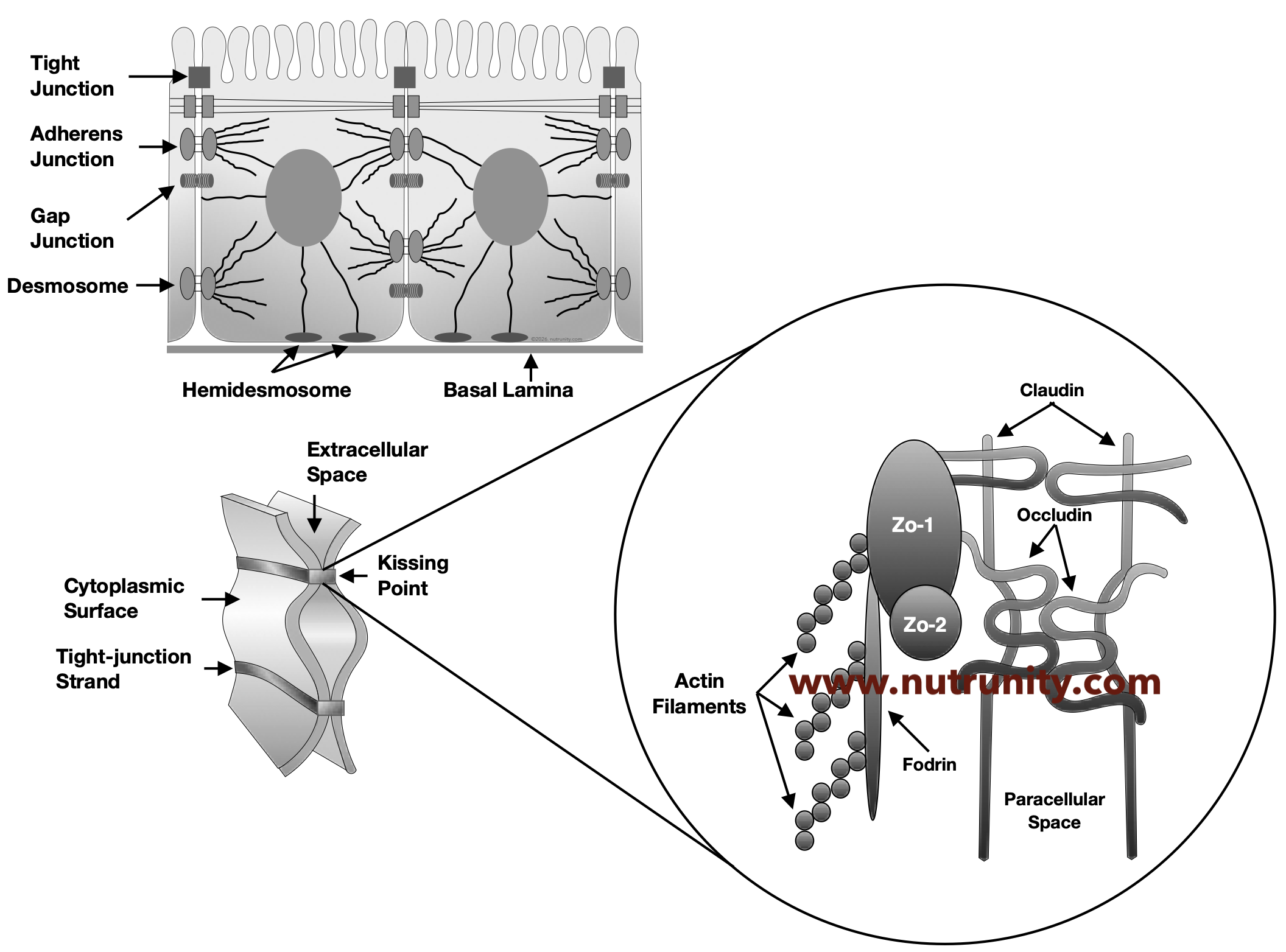
Schematic illustration of tight junctions and the main proteins involved in keeping the gut barrier tight and permeable
What exactly pulls on the seams?
Think of it using the same analogy: Tiles + grout ➡ inflammation ➡ grout dries/cracks + tiles shift. It helps; you can think of it as Velcro. You have a hook-like side and a soft, fluffy opposite side to which it can attach. Inflammation reduces how well the Velcro hooks stick (via protein modification) and sometimes strips some Velcro off (via endocytosis/proteases).
Velcro is very easy to pull apart and put back together. It can also be distorted if not put properly, potentially leaving a gap. Sometimes you can pull too hard, and the seam breaks off, leaving a gap under either layer. The velcro itself is untouched. So, think of it as leaky gut. In all these scenarios, the barrier is no longer tight and permeable, and anything can pass through without restriction.
What can make the seals “loosen” during illness
Tight junctions get “looser” (or break down) when inflammation, microbes/toxins, oxidative injury, or cell damage forces the junction proteins to (a) be pulled apart by cellular contraction, (b) be removed from the membrane by endocytosis, (c) be cut by proteases, (d) be chemically damaged by ROS/NO, and/or (e) disappear because the lining cells die/extrude.
When you have an infection or inflammation, several things can happen at the same time:
1. Inflammation signals make the cells “pull” the junction open (mechanical tension)
Why it happens: the immune system releases alarm chemicals (cytokines) to fight threats, but these signals also change how epithelial cells use their internal “muscle” system.
How it happens (mechanism): Cytokines such as TNF‑α and IL‑1β activate intracellular pathways that increase myosin light chain kinase (MLCK) levels/activity, leading to contractions that physically strain the junction. Other cytokines (IFN‑γ) can also increase contraction through a parallel pathway that increases cytoskeletal tension.
In immune‑mediated colitis, TNF-driven MLCK recruitment to this belt generates force that pulls on ZO‑1 (a key “anchor” protein) and can cause it to detach from the junctional complex. (see illustration 2)
2. Cells “pull junction parts inside” (endocytosis/internalisation)
Why it happens: cells constantly recycle membrane proteins; during inflammation or infection, this recycling can be redirected to remove junction proteins from the cell surface.
How it happens: TNF/IL‑1β/LPS and related signals can trigger occludin endocytosis after MLCK activation. This can also occur while tension is simultaneously disrupting ZO‑1, two hits at once.
3. Microbes (and sometimes our own enzymes) can “cut” junction proteins
Why it happens: Many microbes benefit if they can weaken the barrier; host proteases released during inflammation can also play a decisive part.
How it happens: Some bacterial proteases first loosen tight junctions, then enable toxins to reach and cleave deeper adhesion proteins, collapsing cell–cell adhesion more broadly.
Protease-triggered pathways can also promote junction internalisation/disassembly.
4. Oxidative/nitrosative stress chemically damages junctions
Why it happens: In severe inflammation (e.g., sepsis/critical illness), cells generate reactive oxygen species (ROS) and excess nitric oxide (NO).
How it happens: ROS can oxidise occludin/claudins, causing conformational changes, mislocalisation, and proteasomal degradation, and can also exacerbate inflammatory signalling (MAPK/NF‑κB), which further reduces junctional protein expression and promotes cell death.
Excess NO (from dysbiotic/inflammatory conditions) can attack tight junction proteins, promote apoptosis/necrosis, and impair epithelial repair.
5. Post‑translational modifications weaken the ‘anchors’ that hold junctions in place
Why it happens: Signalling kinases are activated during inflammation/infection and modify junction proteins.
How it happens: Occludin’s binding to scaffold proteins (like ZO‑1) depends on its phosphorylation state; inflammatory/oxidant-activated kinases can reduce ZO‑1 binding, making junctions easier to disassemble.
6. Cell death or extrusion removes the junctions entirely
Why it happens: Severe insults can kill epithelial cells or force them to be extruded; junctions cannot exist without intact adjacent cells. Imagine the intestinal lining as a paper garland made of human shapes holding hands. The “hands” are tight junctions. As long as every human is present, the chain is continuous and intact.
Severe insults can kill epithelial cells or force them to be shed. This is like cutting one or more humans out of the garland with scissors. Where a cell is missing, the junctions literally cannot exist; the chain is broken at that point. You can use Sellotape to “repair” the garland.
Just as Sellotape does not instantly restore the original paper human, the early repair is a temporary patch: functional, but not yet identical to uninjured tissue. Full restoration of cell height, polarity, mucus production and junctional complexity takes more time and cell turnover. The more cells are missing, the longer it takes to repair and the barrier to function effectively once more.
In response to insults, surviving neighbouring cells spread and crawl to cover the gap, and new tight junctions form between them (the biological equivalent of sticking the garland back together with Sellotape). Over the next hours to days, stem cells generate new epithelial cells to replace those that were lost, restoring the lining's full thickness and function.
In health, this repair process is highly efficient. Problems arise when insults are so severe, or so frequent, that the garland is repeatedly cut faster than it can be taped and rebuilt. That is when persistent barrier dysfunction and symptoms are more likely to occur.
How it happens: In sepsis, intestinal epithelial apoptosis increases markedly and correlates with severity; loss of epithelial cells creates gaps that overwhelm normal repair/remodeling.